.png)
)_edited_pn.png)


Why Your MRI Doesn't Match Your Pain (And What Actually Matters More)
The Most Confusing Conversation in Orthopedics
It happens in our office weekly, in both directions:
"My MRI is terrible, but I feel fine." Advanced arthritis, torn meniscus, bulging everything — and the patient came in for something else entirely.
"My MRI is clean, but I'm in agony." Normal imaging, real suffering — and a stack of doctors who've implied the pain must not be real.
Both are completely normal. And both reveal the same truth: the MRI shows anatomy, not pain.
What the Research Actually Shows
The majority of adults over 50 have meniscus tears, disc bulges, or rotator cuff abnormalities on MRI — with zero symptoms. These findings are so common they're essentially wrinkles on the inside.
Conversely, some of the most painful conditions we treat — early tendinopathy, bursitis, nerve entrapments, myofascial pain — can look completely unremarkable on standard MRI.
Pain is generated by the nervous system responding to signals from tissue — inflammation, mechanical irritation, sensitization — most of which don't photograph well.
An MRI is a structural inventory. Pain is a physiologic event. They're related, but they're not the same thing, and treating the picture instead of the patient is one of the most common errors in musculoskeletal care.
The Two Traps
Trap 1: Treating the MRI, not the patient. The scan shows a degenerative meniscus tear, so it gets scoped — even though the pain pattern pointed to arthritis all along. Sham-controlled trials have shown this surgery adds little for degenerative tears. The MRI was real. It just wasn't the problem.
Trap 2: Dismissing pain because the MRI is clean. "Your scan looks fine" is not a diagnosis. It means the answer isn't visible on that particular test. Tendinopathy, bursitis, and nerve irritation are clinical diagnoses — made by history and exam, confirmed by targeted ultrasound, not by MRI.
What Actually Matters More
The story — where it hurts, when, what makes it worse, what injury (if any) started it. A good history outperforms most imaging.
The exam — what reproduces the pain, what's weak, what's unstable. Hands on the patient, not eyes on the screen.
Concordance — does the imaging finding match the symptoms? A medial meniscus tear with medial joint-line pain is concordant. A lateral meniscus tear with medial pain is a bystander.
Dynamic imaging — ultrasound shows the tissue moving in real time, which is how many tendon and nerve problems actually reveal themselves.
Our Rule
We image to confirm or refine a clinical diagnosis — never to go fishing. If your MRI and your pain disagree, we believe the pain and keep looking. The scan is a consultant, not the boss.
Bring Us the Confusing Case
If you've been told "everything looks fine" but you're not fine — or told you need surgery based on a scan alone — get a second opinion. Call Carolina Nonsurgical Orthopedics at 919.719.2270 or schedule online. Offices in Raleigh and Cary, NC. We'll examine you first and image second, in that order.
About Your Physicians
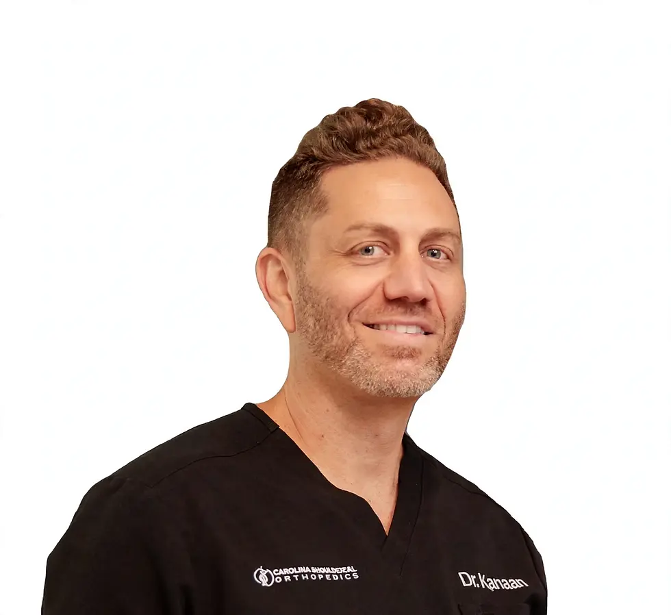
Dr. Matthew Kanaan
Nonsurgical Orthopedic Specialist
Founder of Carolina Nonsurgical Orthopedics. Fellowship trained in sports medicine at Duke; residency in family medicine at Duke, where he served as Chief Resident.
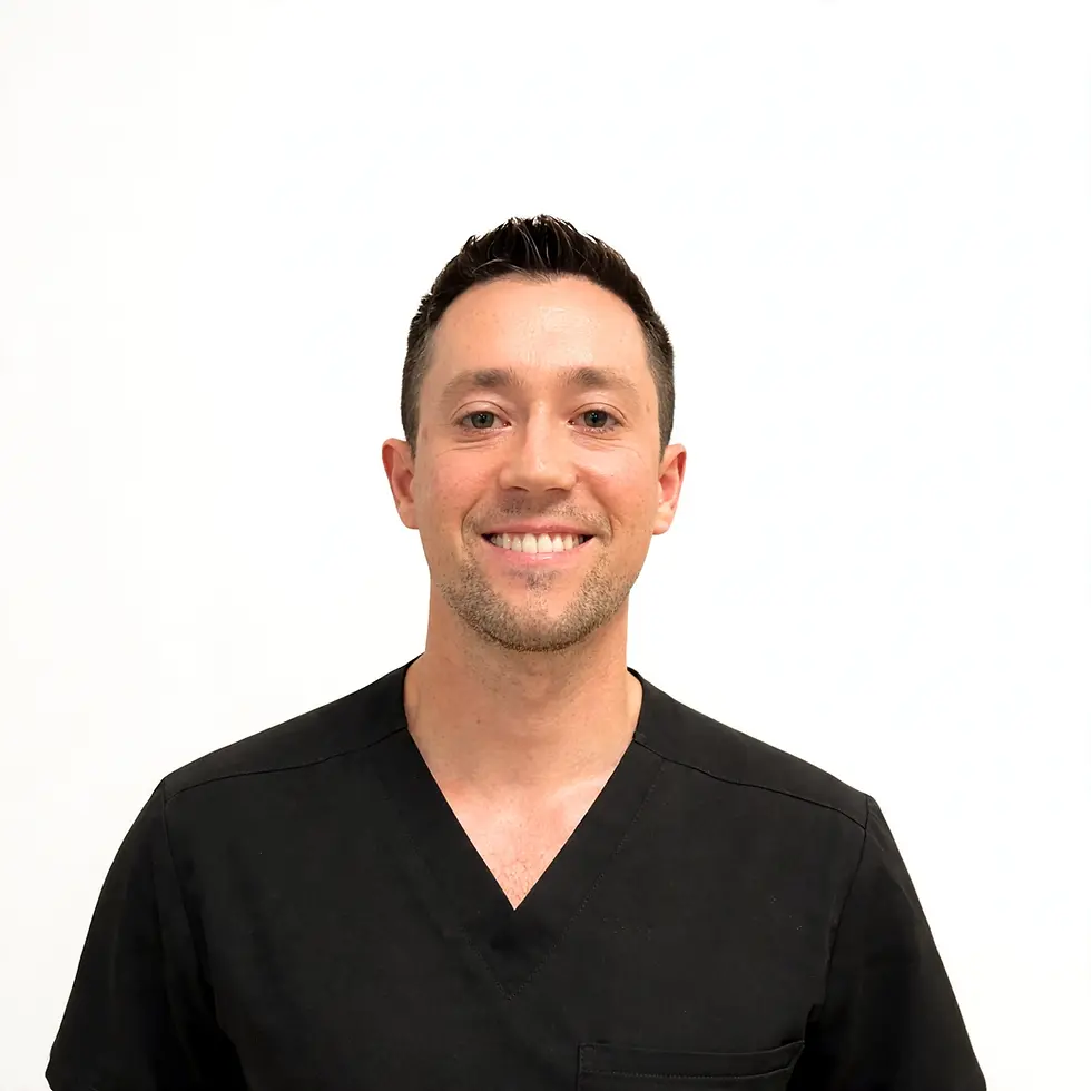
Dr. Pierce Knox
Nonsurgical Orthopedic Specialist
Fellowship trained in nonsurgical sports medicine at Penn Medicine; residency in primary care at Christiana Care.